Home
/ Diagram Of The Muscles In The Forearm : Anatomy Of Human Arm Muscular System Download Scientific Diagram - In these diagrams, the brachioradialis muscle is indicated.
Diagram Of The Muscles In The Forearm : Anatomy Of Human Arm Muscular System Download Scientific Diagram - In these diagrams, the brachioradialis muscle is indicated.
Diagram Of The Muscles In The Forearm : Anatomy Of Human Arm Muscular System Download Scientific Diagram - In these diagrams, the brachioradialis muscle is indicated.. It is a functionally important muscle that contains two heads. Remembering the action of each one can be quite difficult. Anterolateral surface of radius distal to radial tuberosity. Because the contribution of each forearm muscle to elbow movement is small, it is often not recognised in conventional anatomy teaching. In these diagrams, the brachioradialis muscle is indicated.
Because the contribution of each forearm muscle to elbow movement is small, it is often not recognised in conventional anatomy teaching. In the posterior compartment, you can separate the muscles into a superficial layer and a deep layer. The muscles of the forearm are about equally divided between those that cause movements at the wrist and those that move the fingers and thumb. Try labeling diagrams and worksheets as additional learning aids. The antibrachial or forearm muscles may be divided into a volar and a dorsal group.
Muscles Of The Anterior Forearm Flexion Pronation Teachmeanatomy from teachmeanatomy.info Inflammation of this region caused by repetitive. Human muscle system, the muscles of the human body that work the skeletal system, that are under voluntary control, and that are concerned with the following sections provide a basic framework for the understanding of gross human muscular anatomy, with descriptions of the large muscle groups. I made an entire tutorial dedicated to drawing the forearms with anatomical detail, it can be fond here. Click here for access to the full anatomy glossary. The antibrachial or forearm muscles may be divided into a volar and a dorsal group. The forearm is the region of the upper limb between the elbow and the wrist. The forearm is the region of the upper limb between the elbow and the wrist. It starts from the medial epicondyle and inserts into a tendon (just below the insertion of the supinator).
Diagram the movements of the humerus muscles that act on the forearm.
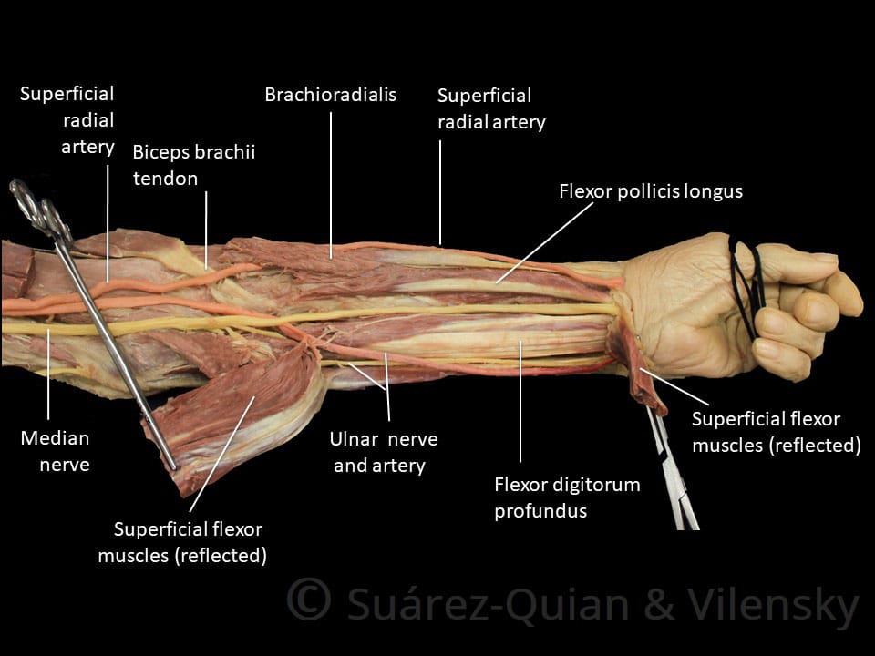
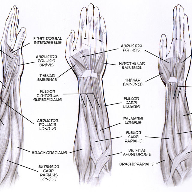
Forearm muscles in the anterior compartment are arranged in superficial, intermediate and deep categories. Inflammation of this region caused by repetitive. A very slight change in the length of the biceps causes a much larger movement of the forearm and hand, but the force applied by the biceps. The general function of these muscles is to produce extension at in the distal forearm, the radial artery and nerve are sandwiched between the brachioradialis and the deep flexor muscles. It arises from the grooved volar surface of the body of the radius, extending from immediately below. Remembering the action of each one can be quite difficult. Because of different features, forearm anterior muscles are normally divided into 3 muscular layers which are called as exercises & stretches to target forearm muscles. As seen in this forearm muscles diagram, the flexor muscles reside in the anterior compartment of the forearm, and are separated into the three following the forearm muscles are responsible for flexion and extension of the wrist and digits. The flexor pollicis longus is situated on the radial side of the forearm, lying in the same plane as the preceding. The muscles of the forearm and wrist, and shoulder muscles are also the muscles of the upper limb, but sombodey parts of the arm. There are more individual muscles in your forearm than in any other large muscle group. In the superficial layer there are four muscles which all arise from a common tendon attached to the medial epicondyle of the humerus, so this attachment site is called the common flexor origin. Superficial muscles of the posterior forearm:
Superficial muscles of the posterior forearm: The antibrachial or forearm muscles may be divided into a volar and a dorsal group. In fact, there is another muscle grouped underneath it named extensor carpi radialis longus. This muscle is part of muscle anatomy master class. Medial epicondyle of humerus i:
Muscles Of The Lower Arm And Hand Human Anatomy And Physiology Lab Bsb 141 from s3-us-west-2.amazonaws.com This is a fusiform muscle that forms the lateral boundary of the cubital fossa and is the most superficial muscle on the radial side of the forearm. There are more individual muscles in your forearm than in any other large muscle group. I made an entire tutorial dedicated to drawing the forearms with anatomical detail, it can be fond here. The flexor pollicis longus is situated on the radial side of the forearm, lying in the same plane as the preceding. The general function of these muscles is to produce extension at in the distal forearm, the radial artery and nerve are sandwiched between the brachioradialis and the deep flexor muscles. Superficial muscles of the posterior forearm: The accompanying muscle diagram reveals the muscles' positions beneath the surface. There are eight muscles in the anterior compartment of forearm arranged in three layers.
I've just switched over to a diagram to show you this muscle.
In these diagrams, the brachioradialis muscle is indicated. A deep layer, intermediate layer and superficial layer. Superficial muscles of the posterior forearm: The pronator teres muscle forms the medial border of the cubital fossa in the anterior elbow. Try labeling diagrams and worksheets as additional learning aids. Lateral epicondyle of humerus and ulna distal to radial notch i: Tutorials and quizzes on muscles that act on the forearm/ forearm muscles (flexors and extensors of the forearm), using interactive animations and diagrams. The forearm is the region of the upper limb between the elbow and the wrist. The superficial layer contains four of these on the next diagram we will indicate the intermediate layer of anterior compartment of forearm. I've just switched over to a diagram to show you this muscle. This layer contains only one muscle, the flexor digitorum. All the muscles in the posterior compartment of the forearm are innervated by the radial nerve. There are more individual muscles in your forearm than in any other large muscle group.
Remembering the action of each one can be quite difficult. The anterior forearm muscles are divided into 3 muscular layers; The forearm is a mass of some 20 different muscles. Lateral epicondyle of humerus and ulna distal to radial notch i: Muscles that participate in the same action, such as flexing the forearm, are actually partitioned off within the body into compartments by a tendinous sheathing called the intermuscular septum.
Forearm Muscle Diagram A Contrast Sketch Of Forearm Muscles With The Download Scientific Diagram from www.researchgate.net Medial epicondyle of humerus i: The muscles of the anterior of the forearm are generally divided into two groups:superficial deepsuperficial muscles of the front of the forearm this group consists of five muscles. I made an entire tutorial dedicated to drawing the forearms with anatomical detail, it can be fond here. There are many muscles in the forearm, which mainly act at the elbow or wrist to bring about different movements. In these diagrams, the brachioradialis muscle is indicated. Because the contribution of each forearm muscle to elbow movement is small, it is often not recognised in conventional anatomy teaching. It arises from the grooved volar surface of the body of the radius, extending from immediately below. Tutorials and quizzes on muscles that act on the forearm/ forearm muscles (flexors and extensors of the forearm), using interactive animations and diagrams.
Lateral epicondyle of humerus and ulna distal to radial notch i:
Diagram of the muscles of the arm in action. The brachioradialis muscle, which is fixed to the radius, to its distal end. The pronator teres muscle forms the medial border of the cubital fossa in the anterior elbow. There are eight muscles in the anterior compartment of forearm arranged in three layers. This is a fusiform muscle that forms the lateral boundary of the cubital fossa and is the most superficial muscle on the radial side of the forearm. I've just switched over to a diagram to show you this muscle. The muscles of the upper arm are responsible for the flexion and extension of the forearm at the elbow joint. Pronator teres pronates the forearm, turning the hand posteriorly. The forearm is the region of the upper limb between the elbow and the wrist. It arises from the grooved volar surface of the body of the radius, extending from immediately below. The forearm is a mass of some 20 different muscles. The term forearm is used in anatomy to distinguish it from the arm. Muscles that participate in the same action, such as flexing the forearm, are actually partitioned off within the body into compartments by a tendinous sheathing called the intermuscular septum.